We are familiar with the concepts of particle and wave in our everyday experience. Marble balls, grains of sand, atoms, electrons and so on are some examples of particles while the examples of waves are sea waves, ripples in a pond, sound waves and light waves.
Particle is a material object which is considered as a tiny concentration of matter (localized in space and time) whereas wave is a broad distribution of energy (not localized in space and time). They, both particles and waves, have the ability to carry energy and momentum from one place to another.
Classical physics which describes the motion of the macroscopic objects treats particles and waves as separate components of physical reality. The mechanics of particles and the optics of waves are traditionally independent subjects, each with its own experiments and principles.
Electromagnetic radiations are regarded as waves because they exhibit wave nature in phenomena such as interference, diffraction and polarization under some suitable circumstances. Similarly, under other circumstances like black body radiation and photo electric effect, electromagnetic radiations behave as though they consist of stream of particles.
When electrons, protons and other particles are discovered, they are considered as particles because they possess mass and charge. However, later experiments showed that under certain circumstances, they exhibit wave- like properties also.
In this unit, the particle nature of waves (radiation) and the wave nature of particles (matter) - that is, wave- particle duality of radiation and matter is discussed with the relevant experimental observations supporting this dual nature.
8.1.1 Electron emission#
In metals, the electrons in the outer most shells are loosely bound to the nucleus. Even at room temperature, there are a large number of free electrons which are moving inside the metal in a random manner. Though they move freely inside the metal, they cannot leave the surface of the metal. The reason is that when free electrons reach the surface of the metal, they are attracted by the positive nuclei of the metal. It is this attractive pull which will not allow free electrons to leave the metallic surface at room temperature.
In order to leave the metallic surface, the free electrons must cross a potential barrier created by the positive nuclei of the metal. The potential barrier which prevents free electrons from leaving the metallic surface is called surface barrier.
Free electrons possess some kinetic energy and this energy is different for different electrons. The kinetic energy of the free electrons is not sufficient to overcome the surface barrier. Whenever an additional energy is given to the free electrons, they will have sufficient energy to cross the surface barrier and they escape from the metallic surface. The liberation of electrons from any surface of a substance is called electron emission.
The minimum energy needed for an electron to escape from the metal surface is called work function of that metal. The work function of the metal is denoted by $\Phi_0$ and is measured in electron volt (eV).
Note The SI unit of energy is joule. But electron volt is a commonly used unit of energy in atomic and nuclear physics.
One electron volt is defined as the kinetic energy gained by an electron when accelerated by a potential difference of 1 V.
$$ 1eV = \mathrm{KE} \text{ gained by the electron } = \text{Work done by the electric field} = qV = 1.602\times 10^{-19}\mathrm{C}\times 1\mathrm{V} = 1.602\times 10^{-19}\mathrm{J} $$Suppose the maximum kinetic energy of the free electron inside the metal is $0.5\mathrm{eV}$ and the energy needed to overcome the surface barrier of a metal is $3\mathrm{eV}$ then the minimum energy needed for electron emission from the metallic surface is $3 - 0.5 = 2.5\mathrm{eV}$. Here $2.5\mathrm{eV}$ is the work function of the metal.
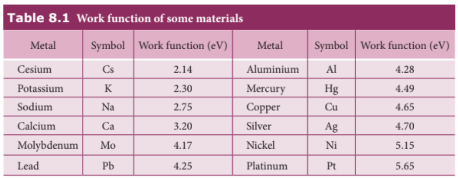
The work function is different for different metals and is a typical property of metals and the nature of their surface. Table 8.1 gives the approximate value of work function for various metals. The material with smaller work function is more effective in electron emission because extra energy required to release the free electrons from the metal surface is smaller.
So the metal selected for electron emission should have low work function. The electron emission is categorized into different types depending upon the form of energy being utilized. There are mainly four types of electron emission which are given below.
i) Thermionic emission#
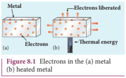
When a metal is heated to a high temperature, the free electrons on the surface of the metal get sufficient energy in the form of thermal energy so that they are emitted from the metallic surface (Figure 8.1). This type of emission is known as thermionic emission.
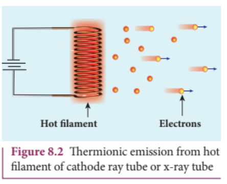
The intensity of the thermionic emission (the number of electrons emitted) depends on the metal used and its temperature. Examples: cathode ray tubes, electron microscopes, x- ray tubes etc (Figure 8.2).
ii) Field emission#
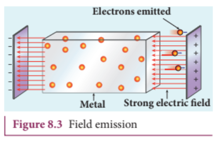
Electric field emission occurs when a very strong electric field is applied across the metal. This strong field pulls the free electrons and helps them to overcome the surface barrier of the metal (Figure 8.3). Examples: Field emission scanning electron microscopes, Field- emission display etc.
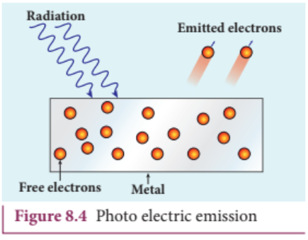
iii) Photo electric emission#
When an electromagnetic radiation of suitable frequency is incident on the surface of the metal, the energy is transferred from the radiation to the free electrons. Hence, the free electrons get sufficient energy to cross the surface barrier and the photo electric emission takes place (Figure 8.4). The number of electrons emitted depends on the intensity of the incident radiation. Examples: Photo diodes, photo electric cells etc.
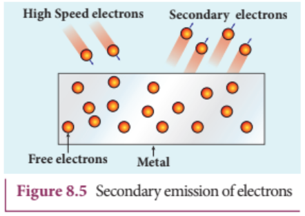
iv) Secondary emission#
When a beam of fast moving electrons strikes the surface of the metal, the kinetic energy of the striking electrons is transferred to the free electrons on the metal surface. Thus the free electrons get sufficient kinetic energy so that the secondary emission of electron occurs (Figure 8.5). Examples: Image intensifiers, photo multiplier tubes etc.
8.2 PHOTO ELECTRIC EFFECT#
8.2.1 Hertz, Hallwachs and Lenard’s observation#
Hertz observation#
Maxwell’s theory of electromagnetism predicted the existence of electromagnetic waves and concluded that light itself is just an electromagnetic wave. Then the experimentalists tried to generate and detect electromagnetic waves through various experiments.
In 1887, Heinrich Hertz was successful in generating and detecting electromagnetic wave with his high voltage induction coil causing a spark discharge between two metallic spheres (we have learnt this in Unit 5 of XII standard physics). When a spark is formed, the charges will oscillate back and forth rapidly and the electromagnetic waves are produced.
The electromagnetic waves thus produced were detected by a detector that has a copper wire bent in the shape of a circle. Although the detection of waves is successful, there is a problem in observing the tiny spark produced in the detector.
In order to improve the visibility of the spark, Hertz made many attempts and finally noticed an important thing that small detector spark became more vigorous when it was exposed to ultraviolet light.
The reason for this behaviour of the spark was not known at that time. Later it was found that it is due to the photoelectric emission. Whenever ultraviolet light is incident on the metallic sphere, the electrons on the outer surface are emitted which caused the spark to be more vigorous.
It is interesting to note that the experiment of Hertz confirmed that light is an electromagnetic wave. But the same experiment also produced the first evidence for particle nature of light.
Hallwachs’ observation#
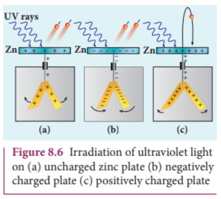
In 1888, Wilhelm Hallwachs, a German physicist, confirmed that the strange behaviour of the spark is due to the action of ultraviolet light with his simple experiment.
A clean circular plate of zinc is mounted on an insulating stand and is attached to a gold leaf electroscope by a wire. When the uncharged zinc plate is irradiated by ultraviolet light from an arc lamp, it becomes positively charged and the leaves will open as shown in Figure 8.6(a).
Further, if the negatively charged zinc plate is exposed to ultraviolet light, the leaves will come closer as the charges leaked away quickly (Figure 8.6(b)). If the plate is positively charged, it becomes more positive upon UV rays irradiation and the leaves open further (Figure 8.6(c)). From these observations, it was concluded that negatively charged electrons were emitted from the zinc plate under the action of ultraviolet light.
Lenard’s observation#
In 1902, Lenard studied this electron emission phenomenon in detail. His simple experimental setup is shown in Figure 8.7. The apparatus consists of two metallic plates $A$ and $C$ placed in an evacuated quartz bulb. The galvanometer $G$ and battery $B$ are connected in the circuit.
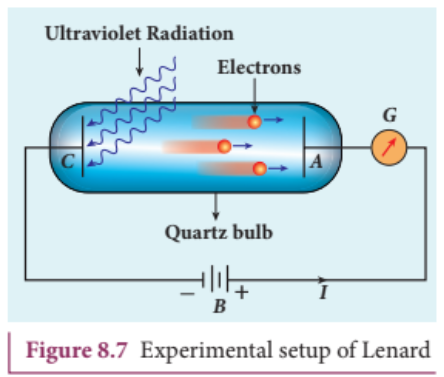
When ultraviolet light is incident on the negative plate $C$ an electric current flows in the circuit that is indicated by the deflection in the galvanometer. On other hand, if the positive plate is irradiated by the ultraviolet light, no current is observed in the circuit.
From these observations, it is concluded that when ultraviolet light falls on the negative plate, electrons are ejected from it which are attracted by the positive plate $A$. On reaching the positive plate through the evacuated bulb, the circuit is completed and the current flows in it. Thus, the ultraviolet light falling on the negative plate causes the electron emission from the surface of the plate.
Photoelectric effect#
The ejection of electrons from a metal plate when illuminated by light or any other electromagnetic radiation of suitable wavelength (or frequency) is called photoelectric effect. Although these electrons are not different from all other electrons, it is customary to call them as photoelectrons and the corresponding current as photoelectric current or photo current.
Metals like cadmium, zinc, magnesium etc show photoelectric emission with ultraviolet light while some alkali metals lithium, sodium, caesium respond well even to larger wavelength radiation like visible light. The materials which eject photoelectrons upon irradiation of electromagnetic wave of suitable wavelength are called photosensitive materials.
8.2.2 Effect of intensity of incident light on photoelectric current#
Experimental setup#
The apparatus shown in Figure 8.8 is employed to study the phenomenon of photoelectric effect in detail. $S$ is a source of electromagnetic waves of known and variable frequency $\nu$ and intensity $I$. $C$ is the cathode (negative electrode) made up of photosensitive material and is used to emit electrons. The anode (positive electrode) $A$ collects the electrons emitted from $C$. These electrodes are kept in an evacuated glass envelope with a quartz window that permits the passage of ultraviolet and visible light.
The necessary potential difference between $C$ and $A$ is provided by high tension battery $B$ which is connected across a potential divider arrangement $PQ$ through a key $K$. $C$ is connected to the centre terminal while $A$ to the sliding contact $J$ of the potential divider. The plate $A$ can be maintained at a desired positive or negative potential with respect to $C$. To measure both positive and negative potential of $A$ with respect to $C$, the voltmeter is designed to have its zero marking at the centre and is connected between $A$ and $C$. The current is measured by a micro ammeter $\mu A$ connected in series.
If there is no light falling on the cathode $C$, no photoelectrons are emitted and the microammeter reads zero. When ultraviolet or visible light is allowed to fall on $C$, the photoelectrons are liberated and are attracted towards anode. As a result, the photoelectric current is set up in the circuit which is measured using micro ammeter.
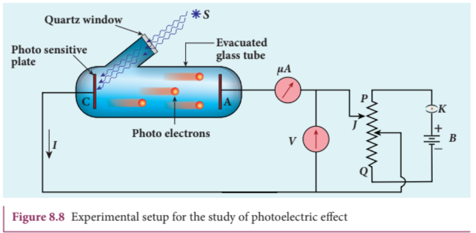
The variation of photocurrent with respect to (i) intensity of incident light (ii) the potential difference between the electrodes (iii) the nature of the material and (iv) frequency of incident light can be studied with the help of this arrangement.
Effect of intensity of incident light on photoelectric current#
To study the effect of intensity of incident light on photoelectric current, the frequency of the incident light and the accelerating potential $V$ of the anode are kept constant. Here the potential of $A$ is kept positive with respect to that of $C$ so that the electrons emitted from $C$ are attracted towards $A$. Now, the intensity of the incident light is varied and the corresponding photoelectric current is measured.
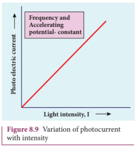
A graph is drawn between light intensity along x- axis and the photocurrent along y- axis. From the graph in Figure 8.9, it is evident that photocurrent - the number of electrons emitted per second - is directly proportional to the intensity of the incident light.
8.2.3 Effect of potential difference on photoelectric current#
To study the effect of potential difference $V$ between the electrodes on photoelectric current, the frequency and intensity of the incident light are kept constant. Initially the potential of $A$ is kept positive with respect to $C$ and the cathode is irradiated with the given light.
Now, the potential of $A$ is increased and the corresponding photocurrent is noted. As the potential of $A$ is increased, photocurrent also increases. However a stage is reached where photocurrent reaches a saturation value (saturation current) at which all the photoelectrons from $C$ are collected by $A$. This is represented by the flat portion of the graph between potential of $A$ and photocurrent (Figure 8.10).
When a negative (retarding) potential is applied to $A$ with respect to $C$, the current does not immediately drop to zero because the photoelectrons are emitted with some definite and different kinetic energies. The kinetic energy of some of the photoelectrons is such that they could overcome the retarding electric field and reach the electrode $A$.
When the negative (retarding) potential of $A$ is gradually increased, the photocurrent starts to decrease because more and more photoelectrons are being repelled away from reaching the electrode $A$. The photocurrent becomes zero at a particular negative potential $V_0$, called stopping or cut- off potential.
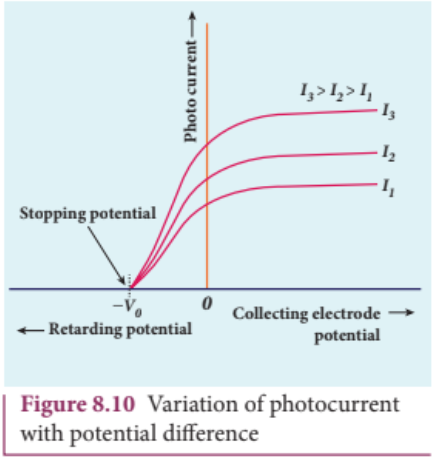
Stopping potential is that value of the negative (retarding) potential given to the collecting electrode $A$ which is just sufficient to stop the most energetic photoelectrons emitted and make the photocurrent zero.
At the stopping potential, even the most energetic electron is brought to rest. Therefore, the initial kinetic energy of the fastest electron $(K_{\mathrm{max}})$ is equal to the work done by the stopping potential to stop it $(eV_0)$.
$$ K_{\mathrm{max}} = \frac{1}{2} m v_{\mathrm{max}}^{2} = eV_0 \quad (8.1) $$where $v_{\mathrm{max}}$ is the maximum speed of the emitted photoelectron.
$$ \begin{array}{l} v_{\mathrm{max}} = \sqrt{\frac{2eV_0}{m}} \\ v_{\mathrm{max}} = \sqrt{\frac{2\times1.602\times10^{-19}}{9.1\times10^{-31}}}\times \sqrt{V_0} \\ = 5.93\times 10^5 \sqrt{V_0} \end{array} \quad (8.2) $$From equation (8.1),
$$ K_{\mathrm{max}} = eV_0 \text{ (in joule)} \quad \text{or} \quad (8.3) $$$$ K_{\mathrm{max}} = V_0 \text{ (in eV)} \quad (8.4) $$From the Figure 8.10, when the intensity of the incident light alone is increased, the saturation current also increases but the value of $V_0$ remains constant.
Thus, for a given frequency of the incident light, the stopping potential is independent of intensity of the incident light. This also implies that the maximum kinetic energy of the photoelectrons is independent of intensity of the incident light.
8.2.4 Effect of frequency of incident light on stopping potential#
To study the effect of frequency of incident light on stopping potential, the intensity of the incident light is kept constant. The variation of photocurrent with the collecting electrode potential is studied for radiations of different frequencies and a graph drawn between them is shown in Figure 8.11. From the graph, it is clear that stopping potential vary over different frequencies of incident light.
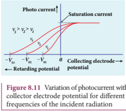
Greater the frequency of the incident radiation, larger is the corresponding stopping potential. This implies that as the frequency is increased, the photoelectrons are emitted with greater kinetic energies so that the retarding potential needed to stop the photoelectrons is also greater.
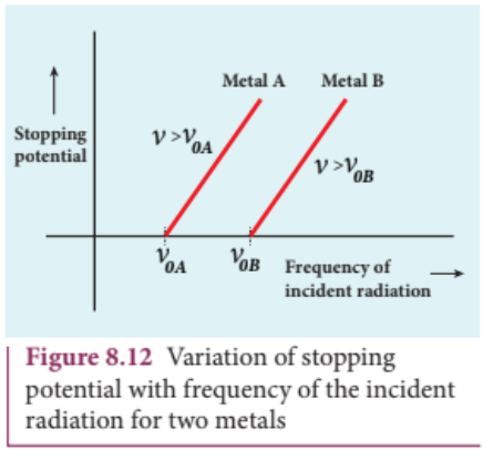
Now a graph is drawn between frequency of incident radiation and the stopping potential for different metals (Figure 8.12). From this graph, it is found that stopping potential varies linearly with frequency. Below a certain frequency called threshold frequency, no electrons are emitted; hence stopping potential is zero for that reason. But as the frequency is increased above threshold value, the stopping potential varies linearly with the frequency of incident light.
8.2.5 Laws of photoelectric effect#
The above detailed experimental investigations of photoelectric effect revealed the following results:
i) For a given metallic surface, the emission of photoelectrons takes place only if the frequency of incident light is greater than a certain minimum frequency called the threshold frequency.
ii) For a given frequency of incident light (above threshold value), the number of photoelectrons emitted is directly proportional to the intensity of the incident light. The saturation current is also directly proportional to the intensity of incident light.
iii) Maximum kinetic energy of the photo electrons is independent of intensity of the incident light.
iv) Maximum kinetic energy of the photo electrons from a given metal is directly proportional to the frequency of incident light.
v) There is no time lag between incidence of light and ejection of photoelectrons.
Once photoelectric phenomenon has been thoroughly examined through various experiments, the attempts were made to explain it on the basis of wave theory of light.
8.2.6 Concept of quantization of energy#
Failures of classical wave theory#
From Maxwell’s theory (Refer unit 5 of volume 1), we learnt that light is an electromagnetic wave consisting of coupled electric and magnetic oscillations that move with the speed of light and exhibit typical wave behaviour. Let us try to explain the experimental observations of photoelectric effect using wave picture of light.
i) When light is incident on a metallic surface, there is a continuous supply of energy to the electrons in the metal surface. According to wave theory, light of greater intensity should impart greater kinetic energy to the liberated electrons (Here, Intensity of light is the energy delivered per unit area per unit time). But this does not happen. The experiments show that maximum kinetic energy of the photoelectrons emitted does not depend on the intensity of the incident light.
ii) According to wave theory, if a sufficiently intense beam of light is incident on the surface, electrons should be liberated from the surface of the target, however low the frequency of the radiation is. From the experiments, it is found that photoelectric emission is not possible below a certain minimum frequency of incident radiation. Therefore, the wave theory fails to explain the existence of threshold frequency.
iii) Since the energy of light is spread across the entire wavefront, the electrons which receive energy from it are large in number. Each electron needs considerable amount of time (a few hours) to get energy sufficient to overcome the work function and to get liberated from the surface. But experiments show that photoelectric emission is almost instantaneous process (the time lag is less than $10^{-9}$ s after the surface is illuminated) which could not be explained by wave theory.
Thus, the experimental observations of photoelectric emission could not be explained on the basis of the wave theory of light.
EXAMPLE 8.1#
For the photoelectric emission from cesium, show that wave theory predicts that
i) maximum kinetic energy of the photoelectrons $(K_{\mathrm{max}})$ depends on the intensity $I$ of the incident light
ii) $K_{\mathrm{max}}$ does not depend on the frequency of the incident light and
iii) the time interval between the incidence of light and the ejection of photoelectrons is very long.
For the sake of simplicity, the following standard assumptions can be made when light is incident on the given material.
a) Light is absorbed in the top atomic layer of the metal b) For a given element, each atom absorbs an equal amount of energy and this energy is proportional to its cross-sectional area $A$ c) Each atom gives this energy to one of the electrons.
Given : The work function for cesium is $2.14\mathrm{eV}$ and the power absorbed per unit area is $1.60\times 10^{-6}\mathrm{Wm}^{-2}$ which produces a measurable photocurrent in cesium.)
Solution
i) According to wave theory, the energy in a light wave is spread out uniformly and continuously over the wavefront.
The energy absorbed by each electron in time $t$ is given by
$$ E = IAt $$With this energy absorbed, the most energetic electron is released with $K_{\mathrm{max}}$ by overcoming the surface energy barrier or work function $\phi_0$ and this is expressed as
$$ K_{\mathrm{max}} = IAt - \phi_0 \quad (1) $$Thus, wave theory predicts that for a unit time, at low light intensities when $IA < \phi_0$, no electrons are emitted. At higher intensities, when $IA \geq \phi_0$, electrons are emitted. This implies that higher the light intensity, greater will be $K_{\mathrm{max}}$.
$K_{\mathrm{max}}$ is dependent only on the intensity under given conditions - that is, by suitably increasing the intensity, one can produce photoelectric effect even if the frequency is less than the threshold frequency. So the concept of threshold frequency does not even exist in wave theory.
ii) According to wave theory, the intensity of a light wave is proportional to the square of the amplitude of the electric field $(E_0^2)$. The amplitude of this electric field increases with increasing intensity and imparts an increasing acceleration and kinetic energy to an electron.
Now $I$ is replaced with a quantity proportional to $E_0^2$ in equation (1). This means that $K_{\mathrm{max}}$ should not depend at all on the frequency of the classical light wave which again contradicts the experimental results.
iii) If an electron accumulates light energy just enough to overcome the work function, then it is ejected out of the atom with zero kinetic energy. Therefore, from equation (1),
$$ 0 = IAt - \phi_0 $$$$ t = \frac{\phi_0}{IA} = \frac{\phi_0}{I(\pi r^2)} $$By taking the atomic radius $r = 1.0\times 10^{-10}\mathrm{m}$ and substituting the given values of $I$ and $\phi_0$, we can estimate the time interval as
$$ t = \frac{2.14\times 1.6\times 10^{-19}}{1.60\times 10^{-6}\times 3.14\times(1\times 10^{-10})^2} $$$$ = 0.68\times 10^{7}s \approx 79 \text{ days} $$Thus, wave theory predicts that there is a large time gap between the incidence of light and the ejection of photoelectrons but the experiments show that photo emission is an instantaneous process.
Concept of quantization of energy#
Max Planck proposed quantum concept in 1900 in order to explain the thermal radiations emitted by a black body and the shape of its radiation curves.
According to Planck, matter is composed of a large number of oscillating particles (atoms) which vibrate with different frequencies. Each atomic oscillator - which vibrates with its characteristic frequency - emits or absorbs electromagnetic radiation of the same frequency. It also says that
i) If an oscillator vibrates with frequency $\nu$ its energy can have only certain discrete values, given by the equation.
$$ E_n = nh\nu \qquad n = 1,2,3\dots \qquad (8.5) $$where $h$ is a constant, called Planck’s constant.
ii) The oscillators emit or absorb energy in small packets or quanta and the energy of each quantum is $h\nu$
This implies that the energy of the oscillator is quantized - that is, energy is not continuous as believed in the wave picture. This is called quantization of energy.
8.2.7 Particle nature of light: Einstein’s explanation#
Einstein extended Planck’s quantum concept to explain the photoelectric effect in 1905. According to Einstein, the energy in light is not spread out over wavefronts but is concentrated in small packets or energy quanta. Therefore, light (or any other electromagnetic waves) of frequency $\nu$ from any source can be considered as a stream of quanta and the energy of each light quantum is given by $E = h\nu$.
He also proposed that a quantum of light has linear momentum and the magnitude of that linear momentum is $p = \frac{h\nu}{c}$. The individual light quantum of definite energy and momentum can be associated with a particle. The light quantum can behave as a particle and this is called photon. Therefore, photon is nothing but particle manifestation of light.
Characteristics of photons:#
According to particle nature of light, photons are the basic constituents of any radiation and possess the following characteristic properties:
i) The photons of light of frequency $\nu$ and wavelength $\lambda$ will have energy, given by
$$ E = h\nu = \frac{hc}{\lambda}. $$ii) The energy of a photon is determined by the frequency of the radiation and not by its intensity and the intensity has no relation with the energy of the individual photons in the beam.
iii) The photons travel with the speed of light and its momentum is given by
$$ p = \frac{h}{\lambda} = \frac{h\nu}{c} $$iv) Since photons are electrically neutral, they are unaffected by electric and magnetic fields.
v) When a photon interacts with matter (photon- electron collision), the total energy, total linear momentum and angular momentum are conserved. Since photon may be absorbed or a new photon may be produced in such interactions, the number of photons may not be conserved.
Note According to quantum concept, intensity of light of given wavelength is defined as the number of energy quanta or photons incident per unit area per unit time, with each photon having same energy. Its unit is $\mathrm{Wm}^{-2}$.
Einstein’s explanation of photoelectric equation#
When a photon of energy $h\nu$ is incident on a metal surface, it is completely absorbed by a single electron and the electron is ejected. In this process, a part of the photon energy is used in overcoming the potential barrier of the metal surface (photoelectric work function $\phi_0$) and the remaining energy as the kinetic energy of the ejected electron. From the law of conservation of energy,
$$ h\nu = \phi_0 + \frac{1}{2} mv^2 \quad (8.6) $$where $m$ is the mass of the electron and $v$ its velocity. This is shown in Figure 8.13(a).
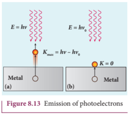
If we reduce the frequency of the incident light, the speed or kinetic energy of photo electrons is also reduced. At some frequency $\nu_0$ of incident radiation, the photo electrons are just ejected with almost zero kinetic energy (Figure 8.13(b)). Then the equation (8.6) becomes
$$ h\nu_0 = \phi_0 $$where $\nu_0$ is the threshold frequency. By rewriting the equation (8.6), we get
$$ h\nu = h\nu_0 + \frac{1}{2} mv^2 \quad (8.7) $$The equation (8.7) is known as Einstein’s photoelectric equation.
If the electron does not lose energy by internal collisions, then it is emitted with maximum kinetic energy $K_{\mathrm{max}}$. Then
$$ K_{\mathrm{max}} = \frac{1}{2} mv_{\mathrm{max}}^2 $$where $v_{\mathrm{max}}$ is the maximum velocity of the electron ejected. The equation (8.6) is rearranged as follows:
$$ K_{\mathrm{max}} = h\nu - \phi_0 \quad (8.8) $$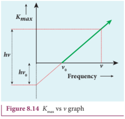
A graph between maximum kinetic energy $K_{\mathrm{max}}$ of the photoelectron and frequency $\nu$ of the incident light is a straight line as shown in Figure 8.14. The slope of the line is $h$ and its $y$-intercept is $-\phi_0$.
Einstein’s equation was experimentally verified by R.A. Millikan. He drew $K_{\mathrm{max}}$ versus $\nu$ graph for many metals (cesium, potassium, sodium and lithium) as shown in Figure 8.15 and found that the slope is independent of the metals.
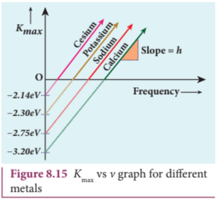
Millikan also calculated the value of Planck’s constant $(h = 6.626\times 10^{-34}\mathrm{Js})$ and work function of many metals (Cs, K, Na, Ca); these values are in agreement with the theoretical prediction.
Explanation for the photoelectric effect:#
The experimentally observed facts of photoelectric effect can be explained with the help of Einstein’s photoelectric equation.
i) As each incident photon liberates one electron, then the increase of intensity of the light (the number of photons per unit area per unit time) increases the number of electrons emitted thereby increasing the photocurrent. The same has been experimentally observed.
ii) From $K_{\mathrm{max}} = h\nu - \phi_0$ it is evident that $K_{\mathrm{max}}$ is proportional to the frequency of the incident light and is independent of intensity of the light.
iii) As given in equation (8.7), there must be minimum energy (equal to the work function of the metal) for incident photons to liberate electrons from the metal surface. Below this value of energy, emission of electrons is not possible. Correspondingly, there exists minimum frequency called threshold frequency below which there is no photoelectric emission.
iv) According to quantum concept, the transfer of photon energy to the electrons is instantaneous so that there is no time lag between incidence of photons and ejection of electrons.
Thus, the photoelectric effect is explained on the basis of quantum concept of light.
The nature of light: wave - particle duality#
We have learnt that wave nature of light explains phenomena such as interference, diffraction and polarization. Certain phenomena like black body radiation, photoelectric effect can be explained by assigning particle nature to light. Therefore, both theories have enough experimental evidences.
In the past, many scientific theories have been either revised or discarded when they contradicted with new experimental results. Here, two different theories are needed to answer the question: what is nature of light?
It is therefore concluded that light possesses dual nature, that of both particle and wave. It behaves like a wave at some circumstances and it behaves like a particle at some other circumstances.
In other words, light behaves as a wave during its propagation and behaves as a particle during its interaction with matter. Both theories are necessary for complete description of physical phenomena. Hence, the wave nature and quantum nature complement each other.
A reader may find it difficult to understand how light can be both a wave and a stream of particle. This is the case even for great scientist like Albert Einstein.
Einstein once wrote a letter to his friend Michel Besso in 1954 expressing his frustration:
“All these fifty years of conscious brooding have brought me no closer to answer the question, ‘What are light quanta?’ Of course today everyone thinks he knows the answer, but he is deluding himself”.
8.2.8 Photo electric cells and their applications#
Photo cell#
Photo electric cell or photo cell is a device which converts light energy into electrical energy. It works on the principle of photo electric effect. When light is incident on the photosensitive materials, their electric properties will get affected, based on which photo cells are classified into three types. They are
i) Photo emissive cell: Its working depends on the electron emission from a metal cathode due to irradiation of light or other radiations. ii) Photo voltaic cell: Here sensitive element made of semiconductor is used which generates voltage proportional to the intensity of light or other radiations. iii) Photo conductive cell: In this, the resistance of the semiconductor changes in accordance with the radiant energy incident on it.
In this section, we discuss about photo emissive cell and its applications.
Photo emissive cell#
Construction:#
It consists of an evacuated glass or quartz bulb in which two metallic electrodes - that is, a cathode and an anode are fixed as shown in Figure 8.16.
The cathode $C$ is semi-cylindrical in shape and is coated with a photo sensitive material. The anode $A$ is a thin rod or wire kept along the axis of the semi-cylindrical cathode. A potential difference is applied between the anode and the cathode through a galvanometer $G$.
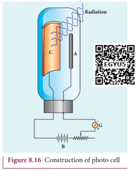
Working:#
When cathode is irradiated with suitable radiation, electrons are emitted from it. These electrons are attracted by anode and hence a current is produced which is measured by the galvanometer. For a given cathode, the magnitude of the current depends on i) the intensity of incident radiation and ii) the potential difference between anode and cathode.
Applications of photo cells:#
Photo cells have many applications, especially as switches and sensors. Automatic lights that turn on when it gets dark use photocells, and street lights that switch on and switch off according to whether it is night or day use photocells.
Photo cells are used for reproduction of sound in motion pictures and are used as timers to measure the speeds of athletes during a race. Photo cells of exposure meters in photography are used to measure the intensity of the given light and to calculate the exact time of exposure.
EXAMPLE 8.2#
A radiation of wavelength $300\mathrm{nm}$ is incident on a silver surface. Will photoelectrons be observed? [work function of silver $= 4.7\mathrm{eV}$]
Solution:
Energy of the incident photon is
$$ E = h\nu = \frac{hc}{\lambda} \text{ (in joules)} $$$$ E = \frac{hc}{\lambda e} \text{ (in eV)} $$Substituting the known values, we get
$$ E = \frac{6.626\times 10^{-34}\times 3\times 10^{8}}{300\times 10^{-9}\times 1.6\times 10^{-19}} $$$$ E = 4.14\mathrm{eV} $$The work function of silver $= 4.7\mathrm{eV}$. Since the energy of the incident photon is less than the work function of silver, photoelectrons are not observed in this case.
EXAMPLE 8.3#
When light of wavelength $2200\mathrm{\AA}$ falls on Cu, photo electrons are emitted from it. Find (i) the threshold wavelength and (ii) the stopping potential. Given: the work function for Cu is $\phi_0 = 4.65\mathrm{eV}$.
Solution:
i) The threshold wavelength is given by
$$ \lambda_0 = \frac{hc}{\phi_0} = \frac{6.626\times 10^{-34}\times 3\times 10^{8}}{4.65\times 1.6\times 10^{-19}} = 2672\mathrm{\dot{A}} $$ii) Energy of the photon of wavelength $2200\mathrm{\AA}$ is
$$ E = \frac{hc}{\lambda} = \frac{6.626\times 10^{-34}\times 3\times 10^{8}}{2200\times 10^{-10}} = 9.035\times 10^{-19}J = 5.65\mathrm{eV} $$We know that kinetic energy of fastest photo electron is
$$ K_{\mathrm{max}} = h\nu - \phi_0 = 5.65 - 4.65 = 1\mathrm{eV} $$From equation (8.3), $K_{\mathrm{max}} = eV_0$
$$ V_0 = \frac{K_{\mathrm{max}}}{e} = \frac{1\times 1.6\times 10^{-19}}{1.6\times 10^{-19}} $$Therefore, stopping potential $= 1\mathrm{V}$
EXAMPLE 8.4#
The work function of potassium is $2.30\mathrm{eV}$. UV light of wavelength $3000\mathrm{\AA}$ and intensity $2\mathrm{Wm}^{-2}$ is incident on the potassium surface. i) Determine the maximum kinetic energy of the photo electrons ii) If $40%$ of incident photons produce photo electrons, how many electrons are emitted per second if the area of the potassium surface is $2\mathrm{cm}^2$?
Solution
i) The energy of the incident photon is
$$ E = \frac{hc}{\lambda} = \frac{6.626\times 10^{-34}\times 3\times 10^{8}}{3000\times 10^{-10}} $$$$ E = 6.626\times 10^{-19}\mathrm{J} = 4.14\mathrm{eV} $$Maximum KE of the photoelectrons is $K_{\mathrm{max}} = h\nu - \phi_0 = 4.14 - 2.30 = 1.84\mathrm{eV}$
ii) The number of photons reaching the surface per second is
$$ n_p = \frac{I}{E}\times A = \frac{2}{6.626\times 10^{-19}}\times 2\times 10^{-4} = 6.04\times 10^{14} \text{ photons / sec} $$The rate of emission of photoelectrons is
$$ = (0.40)n_p = 0.4\times 6.04\times 10^{14} = 2.416\times 10^{14} \text{ photoelectrons / sec} $$EXAMPLE 8.5#
Light of wavelength $390\mathrm{nm}$ is directed at a metal electrode. To find the energy of electrons ejected, an opposing potential difference is established between it and another electrode. The current of photoelectrons from one to the other is stopped completely when the potential difference is $1.10\mathrm{V}$. Determine i) the work function of the metal and ii) the maximum wavelength of light that can eject electrons from this metal.
Solution
i) The work function is given by
$$ \phi_0 = h\nu - K_{\mathrm{max}} = \frac{hc}{\lambda} - eV_0 \quad \text{since } K_{\mathrm{max}} = eV_0 $$$$ = \left[\frac{6.626\times 10^{-34}\times 3\times 10^{8}}{390\times 10^{-9}}\right] - \left[1.6\times 10^{-19}\times 1.10\right] $$$$ = 5.10\times 10^{-19} - 1.76\times 10^{-19} = 3.34\times 10^{-19}\mathrm{J} = 2.09\mathrm{eV} $$ii) The threshold wavelength is
$$ \lambda_0 = \frac{hc}{\phi_0} = \frac{6.626\times 10^{-34}\times 3\times 10^{8}}{3.34\times 10^{-19}} = 5.951\times 10^{-7}\mathrm{m} = 5951\mathrm{\AA} $$8.3 MATTER WAVES#
8.3.1 Introduction - Wave nature of particles#
So far, we learnt that the characteristics of particles and waves are different. A wave is specified by its frequency, wavelength, wave velocity, amplitude and intensity. It spreads out and occupies a relatively large region of space. A particle specified by its mass, velocity, momentum and energy occupies a definite position in space and is very small in size.
Classical physics treated particles and waves as distinct entities. But quantum theory suggested dual character for radiations - that is, radiation behaves as a wave at times and as a particle at other times.
From this wave - particle duality of radiation, the concept of wave nature of matter arises which we will see in this section.
De Broglie wave:#
The wave- particle duality of radiation was extended to matter by a French physicist Louis de Broglie (pronounced as de Broy) in 1924.
Greatly influenced by the symmetry in nature, de Broglie suggested that if radiation like light can act as particles at times, then material particles like electrons can also act as waves at times.
According to de Broglie hypothesis, all material particles like electrons, protons, neutrons in motion are associated with waves. These waves are called de Broglie waves or matter waves.
8.3.2 De Broglie wave length:#
The momentum of photon of frequency $\nu$ is given by
$$ p = \frac{h\nu}{c} = \frac{h}{\lambda} \qquad \text{since } c = \nu \lambda $$The wavelength of a photon in terms of its momentum is
$$ \lambda = \frac{h}{p} \quad (8.9) $$According to de Broglie, the above equation is completely a general one and this is applicable to material particles as well. Therefore, for a particle of mass $m$ travelling with speed $v$, the wavelength is given by
$$ \lambda = \frac{h}{mv} = \frac{h}{p} \quad (8.10) $$This wavelength of the matter waves is known as de Broglie wavelength. This equation relates the wave character (the wave length $\lambda$) and the particle character (the momentum $p$) through Planck’s constant.
8.3.3 De Broglie wave length of electrons:#
Let an electron of mass $m$ be accelerated through a potential difference of $V$ volt. The kinetic energy acquired by the electron is given by
$$ \frac{1}{2} mv^2 = eV $$Therefore, the speed $v$ of the electron is
$$ v = \sqrt{\frac{2eV}{m}} \quad (8.11) $$Hence, the de Broglie wavelength of the matter waves associated with electron is
$$ \lambda = \frac{h}{mv} = \frac{h}{\sqrt{2emV}} $$Substituting the known values in the above equation, we get
$$ \lambda = \frac{6.626\times 10^{-34}}{\sqrt{2V\times 1.6\times 10^{-19}\times 9.11\times 10^{-31}}} $$$$ \qquad = \frac{12.27\times 10^{-10}}{\sqrt{V}} \text{ m} \quad \text{(or)} $$$$ \lambda = \frac{12.27}{\sqrt{V}} \text{ Å} $$For example, if an electron is accelerated through a potential difference of $100\mathrm{V}$ then its de Broglie wavelength is $1.227\mathrm{Å}$.
Since the kinetic energy of the electron, $K = eV$, then the de Broglie wavelength associated with electron can be also written as
$$ \lambda = \frac{h}{\sqrt{2mK}} \quad (8.13) $$8.3.4 Davisson - Germer experiment#
Louis de Broglie hypothesis of matter waves was experimentally confirmed by Clinton Davisson and Lester Germer in 1927. They demonstrated that electron beams are diffracted when they fall on crystalline solids. Since crystal can act as a three- dimensional diffraction grating for matter waves, the electron waves incident on crystals are diffracted off in certain specific directions. Figure 8.17 shows a schematic representation of the apparatus for the experiment.
The filament $F$ is heated by a low tension (L.T.) battery. Electrons are emitted from the hot filament by thermionic emission. They are then accelerated due to the potential difference between the filament and the anode aluminium cylinder by a high tension (H.T.) battery. Electron beam is collimated by using two thin aluminium diaphragms and is allowed to strike a single crystal of Nickel.
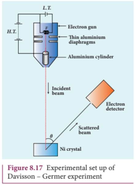
The electrons scattered by Ni atoms in different directions are received by the electron detector which measures the intensity of scattered electron beam. The detector is capable of rotation in the plane of the paper so that the angle $\theta$ between the incident beam and the scattered beam can be changed at our will. The intensity of the scattered electron beam is measured as a function of the angle $\theta$.
It is to be noted that electrons are not the only particles with which wave nature can be demonstrated. The waves are associated with particles like neutrons and alpha particles also when they are in motion. They undergo diffraction when they are scattered by suitable crystals. Neutron diffraction studies are highly useful for investigating crystal structures.
Diffraction is one of the properties of waves. Whenever waves are incident on an obstacle, they bend around the edges of the obstacle. This bending of waves is called diffraction. The amount of bending depends on the wavelength of the waves.
We have learnt in unit 7 that as the wavelength of light is very small, diffraction effects of light are very small. In order to study diffraction of light, diffraction gratings are used. Since x- rays and de Broglie waves of electrons have wavelengths (in the order of $10^{-10}\mathrm{m}$) much shorter than that of the light wave, diffraction grating cannot be used in x- ray diffraction studies. In a crystal, the spacing between atomic planes is comparable to the wavelength of x- rays and de Broglie waves of electrons. Hence, in x- ray diffraction studies, the crystals are used which serve as three- dimensional grating.
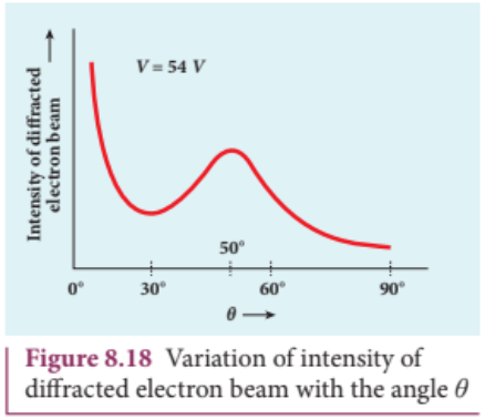
Figure 8.18 shows the variation of intensity of the scattered electrons with the angle $\theta$ for the accelerating voltage of $54\mathrm{V}$. For a given accelerating voltage $V$, the scattered wave shows a peak or maximum at an angle of $50^{\circ}$ to the incident electron beam. This peak in intensity is attributed to the constructive interference of electrons diffracted from various atomic layers of the target material. From the known value of interplanar spacing of Nickel, the wavelength of the electron wave was experimentally calculated as $1.65\mathrm{Å}$.
The wavelength can also be calculated from de Broglie relation for $V = 54\mathrm{V}$ from equation (8.12).
$$ \lambda = \frac{12.27}{\sqrt{V}} \text{ Å} = \frac{12.27}{\sqrt{54}} \text{ Å} $$$$ \lambda = 1.67 \text{ Å} $$This value agrees very well with the experimentally observed wavelength of $1.65\mathrm{Å}$. Thus this experiment directly verifies de Broglie’s hypothesis of the wave nature of moving particles.
8.3.5 Electron Microscope#
Principle#
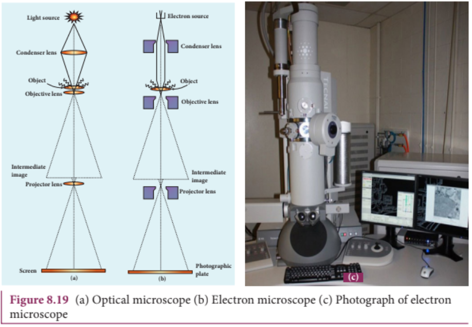
This is the direct application of wave nature of particles. The wave nature of the electron is used in the construction of microscope called electron microscope.
The resolving power of a microscope is inversely proportional to the wavelength of the radiation used for illuminating the object under study. Higher magnification as well as higher resolving power can be obtained by employing the waves of shorter wavelengths.
Louis de Broglie wavelength of electron is very much less than (a few thousands less) that of the visible light being used in optical microscopes. As a result, the microscopes employing de Broglie waves of electrons have very much higher resolving power than optical microscope. Electron microscopes giving magnification more than 2,00,000 times are common in research laboratories.
Working#
The construction and working of an electron microscope is similar to that of an optical microscope except that in electron microscope focussing of electron beam is done by the electrostatic or magnetic lenses. The electron beam passing across a suitably arranged either electric or magnetic fields undergoes divergence or convergence thereby focussing of the beam is done (Figure 8.19).
The electrons emitted from the source are accelerated by high potentials. The beam is made parallel by magnetic condenser lens. When the beam passes through the sample whose magnified image is needed, the beam carries the image of the sample.
With the help of magnetic objective lens and magnetic projector lens system, the magnified image is obtained on the screen. These electron microscopes are being used in almost all branches of science.

EXAMPLE 8.6#
Calculate the momentum and the de Broglie wavelength in the following cases:
i) an electron with kinetic energy $2\mathrm{eV}$ ii) a bullet of $50\mathrm{g}$ fired from rifle with a speed of $200~\mathrm{m / s}$ iii) a $4000\mathrm{kg}$ car moving along the highways at $50~\mathrm{m / s}$
Hence show that the wave nature of matter is important at the atomic level but is not really relevant at macroscopic level.
Solution:
i) Momentum of the electron is
$$ p = \sqrt{2mK} = \sqrt{2\times 9.1\times 10^{-31}\times 2\times 1.6\times 10^{-19}} = 7.63\times 10^{-25} \text{ kg m s}^{-1} $$Its de Broglie wavelength is
$$ \lambda = \frac{h}{p} = \frac{6.626\times 10^{-34}}{7.63\times 10^{-25}} = 0.868\times 10^{-9} \text{ m} = 8.68 \text{ Å} $$ii) Momentum of the bullet is
$$ p = mv = 0.050\times 200 = 10 \text{ kg m s}^{-1} $$Its de Broglie wavelength is
$$ \lambda = \frac{h}{p} = \frac{6.626\times 10^{-34}}{10} = 6.626\times 10^{-35} \text{ m} $$iii) Momentum of the car is
$$ p = mv = 4000\times 50 = 2\times 10^{5} \text{ kg m s}^{-1} $$Its de Broglie wavelength is
$$ \lambda = \frac{h}{p} = \frac{6.626\times 10^{-34}}{2\times 10^{5}} = 3.313\times 10^{-39} \text{ m} $$From these calculations, we notice that electron has significant value of de Broglie wavelength $(\approx 10^{-9}\mathrm{m})$ which can be measured from diffraction studies but moving bullet and car have negligibly small de Broglie wavelengths associated with them $(\approx 10^{-33}\mathrm{m}$ and $10^{-39}\mathrm{m})$ respectively, which are not measurable by any experiment. This implies that the wave nature of matter is important at the atomic level but it is not really relevant at the macroscopic level.
EXAMPLE 8.7#
Find the de Broglie wavelength associated with an alpha particle which is accelerated through a potential difference of $400\mathrm{V}$. Given that the mass of the proton is $1.67\times 10^{-27}\mathrm{kg}$.
Solution
An alpha particle contains 2 protons and 2 neutrons. Therefore, the mass $M$ of the alpha particle is 4 times that of a proton $(m_p)$ (or a neutron) and its charge $q$ is twice that of a proton $(+2e)$.
The de Broglie wavelength associated with it is
$$ \lambda = \frac{h}{\sqrt{2MqV}} = \frac{h}{\sqrt{2\times(4m_p)\times(2e)\times V}} $$$$ = \frac{6.626\times 10^{-34}}{\sqrt{2\times 4\times 1.67\times 10^{-27}\times 2\times 1.6\times 10^{-19}\times 400}} $$$$ = \frac{6.626\times 10^{-34}}{4\times 20\times 10^{-23}\sqrt{1.67\times 1.6}} = 0.00507 \text{ Å} $$EXAMPLE 8.8#
A proton and an electron have same de Broglie wavelength. Which of them moves faster and which possesses more kinetic energy?
Solution
We know that $\lambda = \frac{h}{\sqrt{2mK}}$. Since proton and electron have same de Broglie wavelength, we get
$$ \frac{h}{\sqrt{2m_p K_p}} = \frac{h}{\sqrt{2m_e K_e}} \quad \text{(or)} \quad \frac{K_p}{K_e} = \frac{m_e}{m_p} $$Since $m_e < m_p$, $K_p < K_e$; the electron has more kinetic energy than the proton.
Since $m_e < m_p$, $v_p < v_e$; the electron moves faster than the proton.
8.4 X-RAYS#
Introduction#
Quantum theory of radiation explains photoelectric effect in which the electrons are emitted due to the incidence of photons and the energy is transferred from photons to the electrons. Immediately, a question arises: Is the reverse process also possible?
This means that whether kinetic energy of electron can be transformed into photon energy or not. The phenomenon which answers this question has already been discovered, even before Planck’s quantum theory of radiation.
Discovery of x-rays#
Wilhelm Roentgen in 1895 discovered that whenever fast moving electrons fall on certain materials, a highly penetrating radiation is emitted. Since their origin was not known at that time, they were called x- rays.
X- rays are electromagnetic waves of short wavelength ranging from $0.1$ to $100\dot{A}$. They travel along straight lines with the velocity of light and are not affected by electric and magnetic fields. X- ray photons are highly energetic because of its high frequency or short wavelength. Therefore, they can pass through materials which are opaque to visible light.
The quality of x- rays is measured in terms of their penetrating power which depends on the velocity with which the electrons strike the target material and the atomic number of target material. The intensity of x- rays is dependent on the number of electrons striking the target.
Production of x-rays#
X- rays are produced in x- ray tube which is essentially a discharge tube as shown in Figure 8.20. A tungsten filament $F$ is heated to incandescence by a battery. As a result, electrons are emitted from it by thermionic emission.
The electrons are accelerated to high speeds by the voltage applied between the filament $F$ and the anode. The target materials like tungsten, molybdenum are embedded in the face of the solid copper anode. The face of the target is inclined at an angle of $45^{\circ}$ with respect to the electron beam so that x- rays can leave the tube through its side.
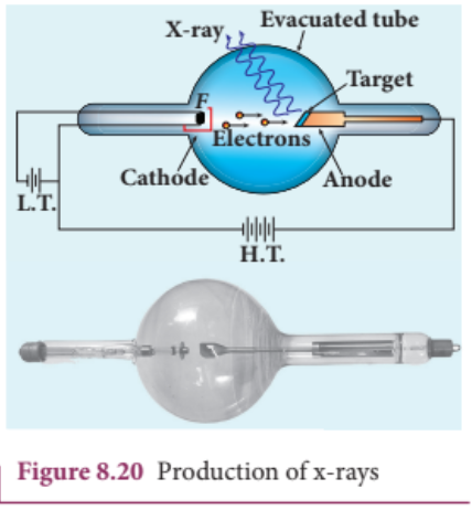
When high- speed electrons strike the target, they are decelerated suddenly and lose their kinetic energy. As a result, x- ray photons are produced. Since most of the kinetic energy of the bombarding electrons gets converted into heat, targets made of high- melting- point metals and a cooling system are usually employed.
X-ray spectra#
X- rays are produced when fast moving electrons strike the metal target. The intensity of the x- rays when plotted against its wavelength gives a curve called x- ray spectrum (Figure 8.21(a) and (b)). X- ray spectra consist of two parts: a continuous spectrum and a series of peaks superimposed on it.
The continuous spectrum consists of radiations of all possible wavelengths with a certain minimum wavelength $\lambda_0$ which depends on the voltage across the electrodes. The peaks are characteristics of the material of the target and hence it is called characteristic spectrum. Figure 8.21(a) depicts the x- ray spectra of tungsten at various accelerating voltages and Figure 8.21(b) shows the x- ray spectra of tungsten and molybdenum at a particular accelerating voltage.
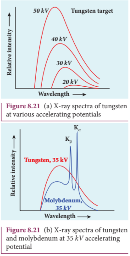
Though classical electromagnetic theory suggests the emission of radiations from accelerating electrons, it could not explain two features exhibited by x- ray spectra. These features are given below.
(i) For a given accelerating voltage, the lower limit for the wavelength of continuous x-ray spectra is same for all targets. This minimum wavelength is called cut-off wavelength. (ii) The intensity of x-rays is significantly increased at certain well-defined wavelengths as shown in the case of characteristic x-ray spectra for molybdenum (Figure 8.21(b)).
But these two features could be explained on the basis of photon theory of radiation.
Continuous x-ray spectra#
When a fast moving electron penetrates and approaches a target nucleus, the interaction between the electron and the nucleus either accelerates or decelerates it which results in a change of path of the electron. The radiation produced from such decelerating electron is called Bremsstrahlung or braking radiation (Figure 8.22).
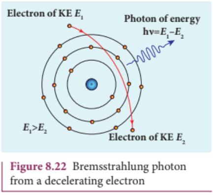
The energy of the photon emitted is equal to the loss of kinetic energy of the electron. Since an electron may lose part or all of its energy to the photon, the photons are emitted with all possible energies (or frequencies). The continuous x- ray spectrum is due to such radiations.
When an electron gives up all its energy, then the photon is emitted with highest frequency $\nu_0$ or lowest wavelength $\lambda_0$. The initial kinetic energy of an electron is given by $eV$ where $V$ is the accelerating voltage. Therefore, we have
$$ h\nu_0 = eV \quad \text{(or)} \quad \frac{hc}{\lambda_0} = eV $$$$ \lambda_0 = \frac{hc}{eV} $$where $\lambda_0$ is the cut- off wavelength. Substituting the known values in the above equation, we get
$$ \lambda_0 = \frac{12400}{V} \text{ Å} \quad (8.14) $$The relation given by equation (8.14) is known as the Duane - Hunt formula.
The value of $\lambda_0$ depends only on the accelerating potential and is same for all targets. This is in good agreement with the experimental results. Thus, the production of continuous x- ray spectrum and the origin of cut - off wavelength can be explained on the basis of photon theory of radiation.
Characteristic x-ray spectra:#
X- ray spectra show some narrow peaks at some well- defined wavelengths when the target is hit by fast electrons. The line spectrum showing these peaks is called characteristic x- ray spectrum. This x- ray spectrum is due to the electronic transitions within the atoms.
When an energetic electron penetrates into the target atom and it can remove some of the $K$- shell electrons. Then the electrons from outer orbits jump to fill up the vacancy so created in the $K$- shell. During the downward transition, the energy difference between the levels is given out in the form of x- ray photon of definite wavelength. Such wavelengths, characteristic of the target, constitute the line spectrum.
From the Figure 8.23, it is evident that $K$- series of lines in the x- ray spectrum of an element arises due to the electronic transitions from $L$, $M$, $N$, … levels to the $K$- level. Similarly, the longer wavelength $L$- series originates when an $L$- electron is knocked out of the atom and the corresponding vacancy is filled by the electronic transitions from $M, N, O$ level to the $L$- level and so on.
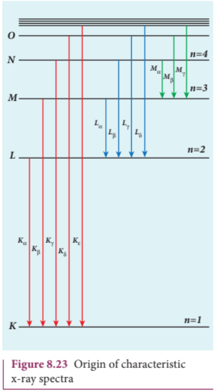
The $K_{\alpha}$ and $K_{\beta}$ of the $K$- series of molybdenum are shown by the two peaks in its x- ray spectrum in Figure 8.21(b).
Applications of x-rays:#
X- rays are being used in many fields. Let us list a few of them.
1) Medical diagnosis#
X- rays can pass through flesh more easily than through bones. Thus an x- ray radiograph containing a deep shadow of the bones and a light shadow of the flesh may be obtained. X- ray radiographs are used to detect fractures, foreign bodies, diseased organs etc.
2) Medical therapy#
Since x- rays can kill diseased tissues, they are employed to cure skin diseases, malignant tumours etc.
3) Industry#
X- rays are used to check for flaws in welded joints, motor tyres, tennis balls and wood. At the custom post, they are used for detection of contraband goods.
4) Scientific research#
X- ray diffraction is important tool to study the structure of the crystalline materials - that is, the arrangement of atoms and molecules in crystals.
EXAMPLE 8.9#
Calculate the cut- off wavelength and cutoff frequency of x- rays from an x- ray tube of accelerating potential 20,000 V.
Solution
The cut- off wavelength of the x- rays in the continuous spectrum is given by
$$ \lambda_{e} = \frac{12400}{V} \text{ Å} = \frac{12400}{20000} \text{ Å} = 0.62 \text{ Å} $$The corresponding frequency is
$$ \nu_0 = \frac{c}{\lambda_0} = \frac{3\times 10^{8}}{0.62\times 10^{-10}} = 4.84\times 10^{18} \text{ Hz} $$Summary#
- $1 \mathrm{eV}$ is equal to $1.602\times 10^{-19}\mathrm{J}$.
- The emission of electrons by supplying thermal energy is known as thermionic emission.
- Electric field emission occurs when a very strong electric field is applied across the metal.
- The emission of electrons due to irradiation of light is called photoelectric emission.
- Secondary emission is the process in which electrons are emitted due to the bombardment of fast moving electrons.
- The photoelectric current (i.e. the number of electrons emitted per second) is directly proportional to the intensity of the incident light.
- Stopping potential is that the value of the negative (retarding) potential given to the collecting electrode $A$ which is just sufficient to stop the most energetic photoelectrons emitted and make the photocurrent zero.
- The stopping potential is independent of intensity of the incident light.
- Maximum kinetic energy of the photoelectrons is independent of intensity of the incident light.
- For a given surface, the emission of photoelectrons takes place only if the frequency of incident light is greater than a certain minimum frequency called the threshold frequency.
- According to Planck, a matter is composed of a large number of oscillating particles (atoms) which vibrate with different frequencies.
- According to Einstein, the energy in light is not spread out over wavefronts but is concentrated in small packets or energy quanta.
- The individual light quantum of definite energy and momentum is called photon.
- Light behaves as a wave during its propagation and behaves as a particle during its interaction with matter.
- Photo electric cell or photo cell is a device which converts light energy into electrical energy.
- According to de Broglie hypothesis, all material particles like electrons, protons, neutrons in motion possess wave nature. These waves associated with them are called de Broglie waves or matter waves.
- Wave nature of the electron is used in the construction of electron microscope.
- Louis de Broglie hypothesis of matter waves was experimentally confirmed by Clinton Davisson and Lester Germer in 1927.
- Whenever fast moving electrons fall on the materials, a highly penetrating radiations, namely x- rays, are emitted.
- Continuous x- ray spectrum consists of radiations of all possible wavelengths with a certain minimum wavelength $\lambda_0$.
- Characteristic x- ray spectra show some narrow peaks at some well- defined wavelengths when the target is hit by fast electrons.
Multiple Choice Questions#
The wavelength $\lambda_{e}$ of an electron and $\lambda_{p}$ of a photon of same energy $E$ are related by (NEET 2013) a) $\lambda_{p}\propto \lambda_{e}$ b) $\lambda_{p}\propto \sqrt{\lambda_{e}}$ c) $\lambda_{p}\propto \frac{1}{\sqrt{\lambda_{e}}}$ d) $\lambda_{p}\propto \lambda_{e}^{2}$
In an electron microscope, the electrons are accelerated by a voltage of $14\mathrm{kV}$. If the voltage is changed to $224\mathrm{kV}$ then the de Broglie wavelength associated with the electrons would a) increase by 2 times b) decrease by 2 times c) decrease by 4 times d) increase by 4 times
The wave associated with a moving particle of mass $3\times 10^{-6}\mathrm{g}$ has the same wavelength as an electron moving with a velocity $6\times 10^{6}\mathrm{m}\mathrm{s}^{-1}$. The velocity of the particle is a) $1.82\times 10^{-18}\mathrm{m}\mathrm{s}^{-1}$ b) $9\times 10^{-2}\mathrm{m}\mathrm{s}^{-1}$ c) $3\times 10^{-31}\mathrm{m}\mathrm{s}^{-1}$ d) $1.82\times 10^{-15}\mathrm{m}\mathrm{s}^{-1}$
When a metallic surface is illuminated with radiation of wavelength $\lambda$ the stopping potential is $V$. If the same surface is illuminated with radiation of wavelength $2\lambda$ the stopping potential is $\frac{V}{4}$. The threshold wavelength for the metallic surface is (NEET 2016) a) $4\lambda$ b) $5\lambda$ c) $\frac{5}{2}\lambda$ d) $3\lambda$
If a light of wavelength $330~\mathrm{nm}$ is incident on a metal with work function $3.55\mathrm{eV}$, the electrons are emitted. Then the wavelength of the wave associated with the emitted electron is (Take $h = 6.6\times 10^{-34}\mathrm{Js}$) a) $< 2.75\times 10^{-9}\mathrm{m}$ b) $\geq 2.75\times 10^{-9}\mathrm{m}$ c) $\leq 2.75\times 10^{-12}\mathrm{m}$ d) $< 2.5\times 10^{-10}\mathrm{m}$
A photoelectric surface is illuminated successively by monochromatic light of wavelength $\lambda$ and $\lambda/2$. If the maximum kinetic energy of the emitted photoelectrons in the second case is 3 times that in the first case, the work function of the material is (NEET 2015) a) $\frac{hc}{\lambda}$ b) $\frac{2hc}{\lambda}$ c) $\frac{hc}{3\lambda}$ d) $\frac{hc}{2\lambda}$
In photoelectric emission, a radiation whose frequency is 4 times threshold frequency of a certain metal is incident on the metal. Then the maximum possible velocity of the emitted electron will be a) $\sqrt{\frac{h\nu_0}{m}}$ b) $\sqrt{\frac{6h\nu_0}{m}}$ c) $2\sqrt{\frac{h\nu_0}{m}}$ d) $\sqrt{\frac{h\nu_0}{2m}}$
Two radiations with photon energies $0.9\mathrm{eV}$ and $3.3\mathrm{eV}$ respectively are falling on a metallic surface successively. If the work function of the metal is $0.6\mathrm{eV}$ then the ratio of maximum speeds of emitted electrons in the two cases will be a) 1:4 b) 1:3 c) 1:1 d) 1:9
A light source of wavelength $520~\mathrm{nm}$ emits $1.04\times 10^{15}$ photons per second while the second source of $460~\mathrm{nm}$ produces $1.38\times 10^{15}$ photons per second. Then the ratio of power of second source to that of first source is a) 1.00 b) 1.02 c) 1.5 d) 0.98
If the mean wavelength of light from sun is taken as $550~\mathrm{nm}$ and its mean power as $3.8\times 10^{26}\mathrm{W}$, then the number of photons emitted per second from the sun is of the order of a) $10^{45}$ b) $10^{42}$ c) $10^{54}$ d) $10^{51}$
The threshold wavelength for a metal surface whose photoelectric work function is $3.313\mathrm{eV}$ is a) $4125\mathrm{Å}$ b) $3750\mathrm{Å}$ c) $6000\mathrm{Å}$ d) $2062.5\mathrm{Å}$
A light of wavelength $500~\mathrm{nm}$ is incident on a sensitive metal plate of photoelectric work function $1.235\mathrm{eV}$. The kinetic energy of the photo electrons emitted is (Take $h = 6.6\times 10^{-34}\mathrm{Js}$) a) $0.58\mathrm{eV}$ b) $2.48\mathrm{eV}$ c) $1.24\mathrm{eV}$ d) $1.16\mathrm{eV}$
Photons of wavelength $\lambda$ are incident on a metal. The most energetic electrons ejected from the metal are bent into a circular arc of radius $R$ by a perpendicular magnetic field having magnitude $B$. The work function of the metal is (KVPY-SX 2016) a) $\frac{hc}{\lambda} - m_e + \frac{e^{2}B^{2}R^{2}}{2m_e}$ b) $\frac{hc}{\lambda} + 2m_e\left[\frac{eBR}{2m_e}\right]^{2}$ c) $\frac{hc}{\lambda} - m_e c^{2} - \frac{e^{2}B^{2}R^{2}}{2m_e}$ d) $\frac{hc}{\lambda} - 2m_e\left[\frac{eBR}{2m_e}\right]^{2}$
The work functions for metals $A$, $B$ and $C$ are $1.92\mathrm{eV}$, $2.0\mathrm{eV}$ and $5.0\mathrm{eV}$ respectively. The metal/metals which will emit photoelectrons for a radiation of wavelength $4100\mathrm{Å}$ is/are a) A only b) both $A$ and $B$ c) all these metals d) none
Emission of electrons by the absorption of heat energy is called………..emission. a) photoelectric b) field c) thermionic d) secondary
Answers
- d
- c
- d
- d
- b
- d
- b
- b
- c
- a
- b
- c
- d
- b
- c
II Short Answer Questions#
- Why do metals have a large number of free electrons?
- Define work function of a metal. Give its unit.
- What is photoelectric effect?
- How does photocurrent vary with the intensity of the incident light?
- Give the definition of intensity of light according to quantum concept and its unit.
- How will you define threshold frequency?
- What is a photo cell? Mention the different types of photocells.
- Write the expression for the de Broglie wavelength associated with a charged particle of charge $q$ and mass $m$, when it is accelerated through a potential $V$.
- State de Broglie hypothesis.
- Why we do not see the wave properties of a baseball?
- A proton and an electron have same kinetic energy. Which one has greater de Broglie wavelength? Justify.
- Write the relationship of de Broglie wavelength $\lambda$ associated with a particle of mass $m$ in terms of its kinetic energy $K$.
- An electron and an alpha particle have same kinetic energy. How are the de Broglie wavelengths associated with them related?
- Define stopping potential.
- What is surface barrier?
- Mention the two features of x- ray spectra, not explained by classical electromagnetic theory.
- What is Bremsstrahlung?
III Long Answer Questions#
- What do you mean by electron emission? Explain briefly various methods of electron emission.
- Briefly discuss the observations of Hertz, Hallwachs and Lenard.
- Explain the effect of potential difference on photoelectric current.
- Explain how frequency of incident light varies with stopping potential.
- List out the laws of photoelectric effect.
- Explain why photoelectric effect cannot be explained on the basis of wave nature of light.
- Give the quantum concept of energy proposed by Max Planck.
- Obtain Einstein’s photoelectric equation with necessary explanation.
- Explain experimentally observed facts of photoelectric effect with the help of Einstein’s explanation.
- Give the construction and working of photo emissive cell.
- Derive an expression for de Broglie wavelength of electrons.
- Briefly explain the principle and working of electron microscope.
- Describe briefly Davisson - Germer experiment which demonstrated the wave nature of electrons.
- List out the characteristics of photons.
- Give the applications photocell.
- How do we obtain characteristic x-ray spectra?
IV. Numerical problems#
How many photons per second emanate from a $50\mathrm{mW}$ laser of $640\mathrm{nm}$? [Ans: $1.61\times 10^{17}\mathrm{s}^{-1}$]
Calculate the maximum kinetic energy and maximum velocity of the photoelectrons emitted when the stopping potential is $81\mathrm{V}$ for the photoelectric emission experiment. [Ans: $1.3\times 10^{-17}\mathrm{J}$, $5.3\times 10^{6}\mathrm{ms}^{-1}$]
Calculate the energies of the photons associated with the following radiation: (i) violet light of $413~\mathrm{nm}$ (ii) X-rays of $0.1\mathrm{nm}$ (iii) radio waves of $10\mathrm{m}$ [Ans: $3\mathrm{eV}$; $12424\mathrm{eV}$; $1.24\times 10^{-7}\mathrm{eV}$]
A $150\mathrm{W}$ lamp emits light of mean wavelength of $5500\mathrm{Å}$. If the efficiency is $12%$, find out the number of photons emitted by the lamp in one second. [Ans: $4.98\times 10^{19}$]
How many photons of frequency $10^{14}$ Hz will make up $19.86$ J of energy? [Ans: $3\times 10^{20}$]
What should be the velocity of the electron so that its momentum equals that of $4000\mathrm{Å}$ wavelength photon. [Ans: $1818\mathrm{ms}^{-1}$]
When a light of frequency $9\times 10^{14}\mathrm{Hz}$ is incident on a metal surface, photoelectrons are emitted with a maximum speed of $8\times 10^{5}\mathrm{ms}^{-1}$. Determine the threshold frequency of the surface. [Ans: $4.61\times 10^{14}\mathrm{Hz}$]
When a $6000\mathrm{Å}$ light falls on the cathode of a photo cell, photoemission takes place. If a potential of $0.8\mathrm{V}$ is required to stop emission of electron, then determine the (i) frequency of the light (ii) energy of the incident photon (iii) work function of the cathode material (iv) threshold frequency and (v) net energy of the electron after it leaves the surface. [Ans: $5\times 10^{14}$ Hz; 2.07 eV; 1.27 eV; $3.07\times 10^{14}$ Hz; 0.8 eV]
A $3310\mathrm{Å}$ photon liberates an electron from a material with energy $3\times 10^{-19}\mathrm{J}$ while another $5000\mathrm{Å}$ photon ejects an electron with energy $0.972\times 10^{-19}\mathrm{J}$ from the same material. Determine the value of Planck’s constant and the threshold wavelength of the material. [Ans: $6.62\times 10^{-34}\mathrm{Js}$; $6620\times 10^{-10}\mathrm{m}$]
At the given point of time, the earth receives energy from sun at $4\mathrm{cal}\ \mathrm{cm}^{-2}\ \mathrm{min}^{-1}$. Determine the number of photons received on the surface of the Earth per $\mathrm{cm}^2$ per minute. (Given : Mean wavelength of sun light $= 5500\mathrm{Å}$) [Ans: $4.65\times 10^{19}$]
UV light of wavelength $1800\mathrm{Å}$ is incident on a lithium surface whose threshold wavelength is $4965\mathrm{Å}$. Determine the maximum energy of the electron emitted. [Ans: $4.40\mathrm{eV}$]
Calculate the de Broglie wavelength of a proton whose kinetic energy is equal to $81.9\times 10^{-15}\mathrm{J}$. (Given: mass of proton is 1836 times that of electron). [Ans: $4\times 10^{-14}\mathrm{m}$]
A deuteron and an alpha particle are accelerated with the same potential. Which one of the two has (i) greater value of de Broglie wavelength associated with it and (ii) less kinetic energy? Explain. [Ans: $\lambda_d = 2\lambda_{\alpha}$ and $K_d = \frac{K_{\alpha}}{2}$]
An electron is accelerated through a potential difference of $81\mathrm{V}$. What is the de Broglie wavelength associated with it? To which part of electromagnetic spectrum does this wavelength correspond? [Ans: $\lambda = 1.36\mathrm{Å}$ and x- rays]
The ratio between the de Broglie wavelength associated with proton, accelerated through a potential of $512\mathrm{V}$ and that of alpha particle accelerated through a potential of $X$ volts is found to be one. Find the value of $X$. [Ans: $64\mathrm{V}$]
References#
- Arthur Beiser, Shobhit Mahajan, Rai Choudhury, Concepts of Modern Physics, Sixth Edition, McGraw Hill Education (India) Private Limited.
- H.S. Mani and G.K. Mehta, Introduction to Modern Physics, Affiliated East-West Press Pvt. Ltd.
- H.C.Verma, Concepts of Physics, Volume 1 and 2, Bharathi Bhawna publishers.
- Halliday, Resnick and Walker, Principles of Physics, Wiley publishers.
ICT CORNER#
Dual nature of radiation and matter#
Topic: Photoelectric effect
In this activity you will be able to visualize how light knocks electrons off a metal target and describe the photoelectric effect experiment.
STEPS:
- Open the browser and type “https://phet.colorado.edu/en/simulation/legacy/photoelectric" in the address bar.
- Change intensity of light and observe how the intensity of light will affect the photo electric current and the energy of electrons.
- By adjusting the value of wavelength and observe how the wavelength of light will affect the photo electric current and the energy of electrons.
- Adjust the value of voltage from the battery and analyse the effect of potential difference on the photoelectric current.
- Change the material of the target and analyse how it will affect the current and the energy of electrons.
- Study the photo electric current - voltage graph and Photo electric current - intensity graph obtained from this experiment.
Note: Install Java application if it is not in your browser. You can download all the phet simulation and works in off line from https://phet.colorado.edu/en/offline-access.
URL: https://phet.colorado.edu/en/simulation/legacy/photoelectric
*Pictures are indicative only. If browser requires, allow Flash Player or Java Script to load the page.